Brain Injury:
In the know
It’s not just your shins that need protection
The brain has a jelly-like consistency and sits within a hard-cased skull; a direct or indirect impact to the head or a whiplash effect will cause the brain to move inside the skull. An impact on the head will involve both linear forces and rotational forces.

Repeated rotational forces cause more brain damage than linear forces.
Rotational Damage
Nature did not design the brain for rotational forces and injury, with the brain being badly designed for sudden rotational acceleration and deceleration. However, in sports, the brain is subjected to sudden acceleration and deceleration injuries. This is because it is rotated by impacts at the side and back of the head. These contacts include:
- Head-to-head;
- Head-to-ball; and
- Head-to-ground.
Head impacts cause damage:
The white and grey matter of the brain is made up of differing densities, which means the jelly-like brain sections move at different speeds when rotated. This causes:
• Unnatural stretching of the long tail (axon) part of brain cells;
• Shearing of brain cells;
• Tearing of fine blood vessels and bleeding;
• Brain cell death; and
• Disruption of the nerve cell connectivity networks in the brain.
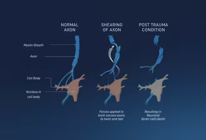 Impacts to the brain can cause damage that is both immediate (axonal damage as shown), and delayed over hours and days (blood flow changes or neural inflammation). This results in Diffuse Axonal Injury (DAI).
DAI is caused by rotational injury to the brain and causes mechanical damage to axons throughout the white matter of the brain. Axons are essential as they allow different parts of the brain to communicate, and are needed for higher intellectual function e.g. controlling behaviours.
DAI is responsible for the majority of cognitive deficits seen after “mild” traumatic brain injury and is particularly responsible for difficulty with thinking, memory and information processing. DAI is not fatal, but the damage caused is cumulative.
Each impact on the brain has the potential to cause an injury that is as visceral as a torn hamstring.
Brain injury happens at the microscopic level, 4,000 times smaller than the eye can see on a regular brain scan.
40% of traumatic brain injuries (the largest group of all brain injuries sustained every year) are classified as mild, in that they don’t register concern based on CT scan results or dictate a need for a CT scan when the individual attends A&E immediately after the accident.
It’s not all about concussion
Research has shown that repetitive sub-concussive impacts (an impact of lower force than required to cause a concussion and without any symptoms) damages the brain, reduces memory and attention, suppresses brain function, contributes to mood and behaviour problems, and leads to neurodegeneration including CTE.
Sub-concussions create an inflammatory response and the production of neurochemicals. This results in damage to the blood-brain barrier, a structure designed to protect the brain. The blood-brain barrier acts as a “security gate”, letting in essential nutrients while keeping harmful toxins and inflammatory cells out. But when damaged or “leaky” it cannot perform this role properly and becomes associated with cognitive decline and neurological damage.
While singular, one-off responses are protective, the problem arises when the brain is subjected to repetitive sub-concussive impacts. The responses from the first injury haven’t yet returned to normal levels, and so subsequent sub-concussive injuries can cause an exaggerated production of neurochemicals and an exaggerated inflammatory response.
This becomes harmful to the brain, rather than protective. It instead damages brain tissue, the blood-brain barrier and leads to the irreversible death of brain cells. Over time, this leads to changes in a brain protein called tau.
So focusing on concussions misses the point of how harmful the accumulation of sub-concussive impacts can be the brain and triggering of longer-term neurodegenerative consequences.
TAU Protein
Tau protein (present in the tiny microtubules of the axons) normally stabilises cognitive brain cells to ensure they communicate and work together efficiently, so an individual can think and behave normally.
When tau protein is damaged from concussive and sub-concussive impacts, it can no longer stabilise the brain cells and they lose their ability to function effectively. The tau protein can also misfold, detach and replicate inside the brain cells with the tau impairing cell function and eventually killing the brain cell. Unfortunately, these brain cells cannot regenerate. The abnormal tau protein also develops the ability to spread to connected brain cells; and once the abnormal tau protein enters a new brain cell, the process starts again, resulting in the death of that brain cell. As tau protein spreads, more of these brain cells – needed for thought, and control of emotions and behaviour – are killed. The symptoms of cognitive impairment and changes in behaviour become increasingly apparent.
The risk and severity of CTE are caused primarily by multiple sub-concussive impacts, and not by one-hit concussions. The force to the brain to cause a concussion is 2-4 times greater than for a sub-concussion, but sub-concussions are over 500 times more frequent [1].
Females are at least three times more seriously impacted by brain trauma than males, with recovery time longer and post-concussive symptoms more pronounced [2].
This is due to:
- Differences in the microstructure of the brain [3], female axons particularly in the white matter tend to be smaller with fewer microtubules, and more likely to be damaged than those of males at the same force application level;
- Hormones and the influence of progesterone on the outcome. In the menstrual cycle there is a significant fluctuation of progesterone levels, whereas males have lower background levels of progesterone;
- Significant differences in head and neck geometry and neck strength in males versus females. Females have a total neck muscle mass to head weight ratio of 1 to 9.36 as compared to a 1 to 3.11 ratio in males; and
- Coaching regimes and the management of injuries.
Consequently, women tend to experience concussion injuries at a lower average impact threshold [4].
Research found that women who were injured during the last two weeks of the menstrual cycle (when progesterone was at its highest) had worse post-concussion symptoms compared with women injured during the first two weeks – when progesterone was low [5]. As hormones fluctuate, grey and white matter volumes change, as does the volume of cerebrospinal fluid. Progesterone is associated with increased tissue and decreased cerebrospinal fluid volume [6].
Females show diminished performance on visual memory and total combined memory function scores after sports-related concussions [7].
Females typically experience intercurrent depression at a rate twice that of males, and tend to experience a more widespread and diverse pattern of post-concussional symptoms [8].
Certain post-concussion symptoms also appear to be more prevalent in women, particularly those of headache, dizziness, fatigue, irritability, and concentration problems three months after sustaining a concussion [9].
It has been 20 years since sex and gender differences were first seen in brain injury research, yet 80.1% of all concussion research data comes from males [10]. This lack of female data means the true extent of damage is likely to be underestimated. Most women, their coaches and medical support are still unaware.
Between ages 8-12 is when peak development of the brain occurs, but the brain is still developing through the refinement and rearrangement of pathways and connections between cells until early 20s.
- Players experiencing head impacts prior to age 12 have been found to have demonstrated worse cognitive, executive (planning, decision-making, impulse control) and new learning abilities as adults, compared to those who were at least 12 years old when they were first exposed to contact and collision sports [11].
- Children and young people who sustain a concussion are at increased risk of developing mental health issues (anxiety and neurotic disorders, behavioural disorders, mood and eating disorders, schizophrenia, substance use disorder and suicidal ideation) [12].
Axons are the tail-like structures that connect neurons in the brain and connect neurons to other cell types. Axons are coated in myelin, and this improves how electrical discharges transfer information along the axon – it is crucial to healthy brain function.
This myelin is formed as the brain develops. It occurs mainly between the second trimester of gestation and the early years of postnatal life, and continues until the mid-20s. Myelin development is linked to cognitive skills in children, as when myelination improves, the brain works faster.
So, children have less myelin than adults. This is problematic, because myelin protects the axon. Axons with little myelin are more exposed and prone to damage from concussions and sub-concussions. Less myelinated axons also don’t recover as well from injury as highly-myelinated axons. This means a child is more vulnerable to brain trauma in sports. Children’s brains undergo noticeable changes after just one season of head impacts, even if they were never diagnosed with a concussion [13].
Research has shown that school pupils and students who experience multiple concussions often struggle with academic performance and learning behaviours long-term [14].
We now know more from research about how the brain responds to rotational forces, than in the past; we know a child’s/youth is more susceptible to these forces. What we don’t know is which child may have a genetic or metabolic risk factor, who will continue to play sport into adulthood and or as a professional, so given the damage of sub-concussions and rotational forces are cumulative, every child is at risk of a massive lifetime dose of sub-concussions and rotational forces. Consequently, their CTE and risk of neurodegenerative disease starts in childhood.
Not Just Part of Professional Sport
Brain injury is not limited to elite or professional sports [15]. The brain does not know if it receives an impact in front of global audiences or in the local park.
Rotational forces from concussive and sub-concussive impacts are a real risk for everyone playing sports, regardless of their position, team, fitness-level and experience.
Whilst concussions may be apparent instantly, the damage from repetitive sub-concussions may not be recognised until years later, meaning it is never too early to protect the brain in sports.
With little pain and no obvious symptoms, these brain injuries remain “hidden in plain sight”. Many players do not even know they need to protect themselves from sub-concussive impacts.

- Leveraging football accelerometer data to quantify associations between repetitive head impacts and chronic traumatic encephalopathy in males, Daneshvar DH, Nair ES, Baucom ZH, Rasch A, Abdolmohammadi B, Uretsky M, Saltiel N, Shah A, Jarnagin J, Baugh CM, Martin BM, Palmisano JN, Cherry JD, Alvarez VE, Huber BR, Weuve J, Nowinski CJ, Cantu RC, Zafonte RD, Dwyer B, Crary JF, Goldstein LE, Kowall NW, Katz DI, Stern RA, Tripodis Y, Stein TD, McClean MD, Alosco ML, McKee AC, Mez J, 2023
- Association of Sex and Age With Mild Traumatic Brain Injury–Related Symptoms: A TRACK-TBI Study, 2021.
- Sex differences in the extent of acute axonal pathologies after experimental concussion, Song H, Tomasevich A, Paolini A, Browne KD, Wofford KL, Kelley B, Kantemneni E, Kennedy J, Qiu Y, Schneider ALC, Dolle JP, Cullen DK, Smith DH, 2024
- Concussive Head Impact Biomechanics in Women’s Lacrosse and Soccer Athletes: A Case Series, Sayre, H.D. et al, 2019.
- Preliminary Report: Localized Cerebral Blood Flow Mediates the Relationship between Progesterone and Perceived Stress Symptoms among Female Collegiate Club Athletes after Mild Traumatic Brain Injury, Yufen Chen, Amy A. Herrold, Virginia Gallagher, Zoran Martinovich, Sumra Bari, Nicole L. Vike, Brian Vesci, Jeffrey Mjaanes, Leanne R. McCloskey, James L. Reilly, Hans C. Breiter, 2021.
- Menstrual cycle-driven hormone concentrations co-fluctuate with white and grey matter architecture changes across the whole brain, Elizabeth J. Rizor, Viktoriya Babenko, Neil M. Dundon, Renee Beverly-Aylwin, Alexandra Stump, Margaret Hayes, Luna Herschenfeld-Catalan, Emily G. Jacobs, Scott T. Grafton, 2023.
- Are there differences in neurocognitive function and symptoms between male and female soccer players after concussions? Covassin, T. et al. 2013.
- Patients with mild traumatic brain injury and acute neck pain at the emergency department are a distinct category within the mTBI spectrum: a prospective multicentre cohort study, Coffeng, S.M. et al. 2020.
- Sex differences in neuropsychological function and post-concussion symptoms of concussed collegiate athletes, Covassin, T., Schatz, P. and Swanik, C.B. 2007
- The International Conference on Concussion in Sport (ICCS), National Athletic Trainers Association (NATA), American Medical Society for Sports Medicine (AMSSM).
- Age of first exposure to football and later-life cognitive impairment in former NFL players, Stamm JM, Bourlas AP, Baugh CM, et al. 2015. 9. Risk of Mental Health Problems in Children and Youths Following Concussion. , Ledoux A, Webster RJ, Clarke AE, et al. 2022
- Subconcussive Head Impact Exposure and White Matter Tract Changes over a Single Season of Youth Football, Naeim Bahrami, Dev Sharma, Scott Rosenthal, Elizabeth M. Davenport, Jillian E. Urban, Benjamin Wagner, Youngkyoo Jung, Christopher G. Vaughan, Gerard A. Gioia, Joel D. Stitzel, Christopher T. Whitlow, and Joseph A. Maldjian, 2016.
- Risk of Mental Health Problems in Children and Youths Following Concussion, Ledoux A, Webster RJ, Clarke AE, et al. 2022.
- Long-Term School Outcomes of Children and Adolescents With Traumatic Brain Injury, Prasad, Mary R. PhD; Swank, Paul R. PhD; Ewing-Cobbs, Linda PhD. 2017.
- Neuropathologic and Clinical Findings in Young Contact Sport Athletes Exposed to Repetitive Head Impacts, McKee AC, Mez J, Abdolmohammadi B, et al. 2023.